43 diagram of a human cell with labels
Diagram Of Body Organs Female Pics Pictures, Images ... - iStock Anatomy of the human body. "r"nBiology education concept. Lateral views. Vector Illustration Male and female reproductive system median section with main parts labeled. Anatomy of the human body. "r"nBiology education concept. Lateral views. Vector Illustration diagram of body organs female pics stock illustrations Diagram of human skin structure — Science Learning Hub Diagram of human skin structure. Add to collection. + Create new collection. Tweet. Rights: University of Waikato Published 1 February 2011 Size: 100 KB Referencing Hub media. The epidermis is a tough coating formed from overlapping layers of dead skin cells.
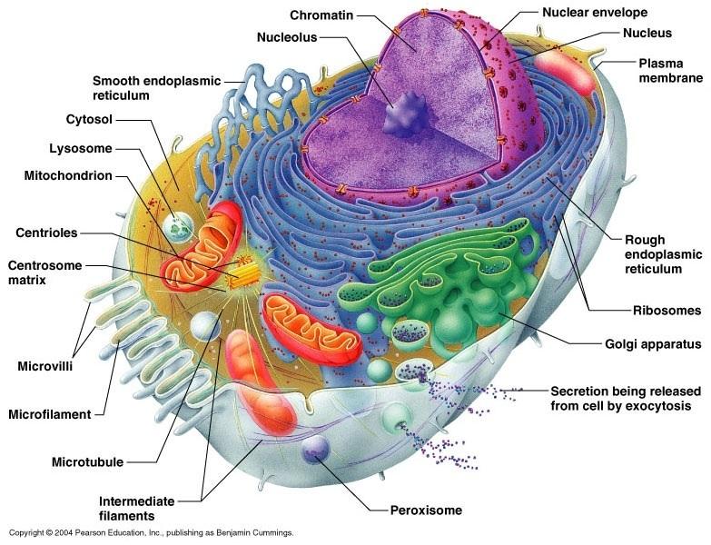
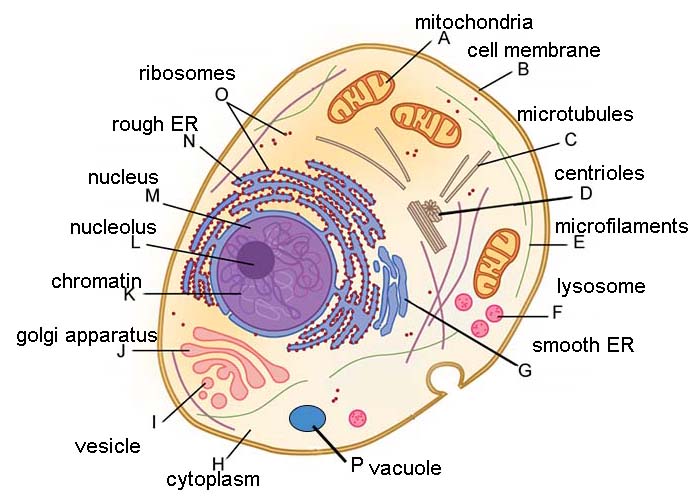
Learn the parts of a cell with diagrams and cell quizzes Labeled cell diagram For this exercise we'll start with an image of a cell diagram ready labeled. Study this and make sure that you're clear about which structure is found where. Cell diagram unlabeled It's time to label the cell yourself!

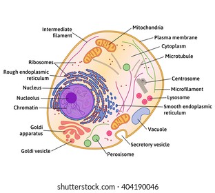
Diagram of a human cell with labels
Cell Diagram | Free Cell Diagram Templates - Edrawsoft A free customizable cells diagram template is provided to download and print. Quickly get a head-start when creating your own cell diagram. Here is a simple cell diagram example created by Science Diagram Maker, which is available in different formats. Cells Diagram | Science Illustration Solutions - Edrawsoft Cells Diagram Symbols Edraw software offers you lots of symbols used in cells diagram like cell structure, paramecium, squamous cell, cell division, bacteria, cell membrane, eggs, sperm, zygote, an animal cell, SARS, tobacco mosaic, adenovirus, coliphage, herpesvirus, AIDS, pollen, plant cell model, onion tissue, etc. Cells Diagram Examples human cell diagram to label Histology basal cell deferens vas label anatomy ansci wisc edu male. Skin histology tissue connective types basic adipose components structure tissues layers system integumentary stain showing dermis dense epidermis irregular embryology. Cell human structure cells cellule nucleus parts anatomy nutshell dna noyau stuff human cell diagram to label
Diagram of a human cell with labels. The Cell - ScienceQuiz.net The diagram shows a plant cell as seen under a microscope. Two of the labels are incorrect. What are they? ... A typical human cell has a diameter of 0.00002 metre ... Liver Diagram with Detailed Illustrations and Clear Labels The liver is one of the most important organs in the human body. Anatomically, the liver is a meaty organ that consists of two large sections called the right and the left lobe. The rib cage partly protects the liver and cannot be felt if you were to touch it. However, it can be felt ascending and descending if you were to take a deep breath. Human Heart Diagram Labeled | Science Trends Daniel NelsonPRO INVESTOR. The human heart is an organ responsible for pumping blood through the body, moving the blood (which carries valuable oxygen) to all the tissues in the body. Without the heart, the tissues couldn't get the oxygen they need and would die. Along with lymphatic vessels, the blood, blood vessels, and lymph, the heart ... Circulatory System Labeled Diagram stock illustrations Browse 154 circulatory system labeled diagram stock illustrations and vector graphics available royalty-free, or start a new search to explore more great stock images and vector art. Newest results Heart Poster Heart blood flow circulation anatomical diagram with atrium and... Anatomy of Nerves of Body and Head
Human Anatomy Label Me! Printouts - EnchantedLearning.com Human Anatomy Label Me! Elementary-level Printouts. Read the definitions then label the diagrams. Advertisement. EnchantedLearning.com is a user-supported site. As a bonus, site members have access to a banner-ad-free version of the site, with print-friendly pages. ... Animal Cell Anatomy Label the animal cell diagram using the glossary of ... Human Body Organs Diagram Stock Photos and Images - Alamy Human body with lymphoid organs (Spleen, Thymus), Lymphatic vessel, lymph nodes, and Cisterna chyli. Vector illustration. ID: 2GD6GGN (RF) Man head and chest anatomy diagram with ghost effect. Skeletal, cardiovascular, nervous and lymphatic systems. ID: T2WN7K (RF) Full length view of male human body with organs. Authentication of Human Cell Lines by STR DNA Profiling ... May 01, 2013 · Since 1951 when the first human cell line, HeLa, was established there has been an increase in the use of human cell lines as models for human diseases such as cancer, substrates for the production of viruses for vaccine production and as tools for the production of recombinant proteins for therapeutics. Unfortunately this accelerated use of human cell lines and the lack of best practices in ... PDF Human Cell Diagram, Parts, Pictures, Structure and Functions Diagram of the human cell illustrating the different parts of the cell. Cell Membrane The cell membraneis the outer coating of the cell and contains the cytoplasm, substances within it and the organelle. It is a double-layered membrane composed of proteins and lipids.
A Well-labelled Diagram Of Animal Cell With Explanation Well-Labelled Diagram of Animal Cell The Cell Organelles are membrane-bound, present within the cells. There are various organelles present within the cell and are classified into three categories based on the presence or absence of membrane. Listed below are the Cell Organelles of an animal cell along with their functions. The Human Skeleton: All You Need to Know - Bodytomy Labeled Skeleton Diagram This skeleton diagram will help explain the different bones of the human body clearly. Cranium The cranium is a skull bone that covers the brain, as seen in the skeleton diagram. The facial bones are not a part of the cranium. The bones that are just above the ear or in front of the ear are known as temporal bones. Stapes Labeled Diagram of the Human Kidney - Bodytomy Labeled Diagram of the Human Kidney The human kidneys house millions of tiny filtration units called nephrons, which enable our body to retain the vital nutrients, and excrete the unwanted or excess molecules as well as metabolic wastes from the body. In addition, they also play an important role in maintaining the water balance of our body. Anatomy (Human Body) Labeling - Exploring Nature Arteries of the Lower Limb (Pelvis, Leg and Foot) Labeling. Arteries of the Upper Limb (Shoulder, Arm, Hand) Labeling. Blood Vessel Anatomy Labeling
Print Chapter 2 cells flashcards | Easy Notecards
ch 8 mastering biology Flashcards | Quizlet The Human Life Cycle 3 of 16 Review Watch this video and then answer the questions. Part A The figure shows the human life cycle. Can you identify the structures and processes? Drag the labels to their appropriate locations on the figure. Pink labels represent structures, and blue labels represent processes.
Ch 3 Review Guide Answer Key
Animal Cell Diagram with Label and Explanation: Cell ... - Collegedunia Animal cell is a typical Eukaryotic cell enclosed by a plasma membrane containing nucleus and organelles which lack cell walls, unlike all other Eukaryotic cells. The typical cell ranges in size between 1-100 micrometers. The lack of cell walls enabled the animal cells to develop a greater diversity of cell types.

1000+ images about Apologia Human Anatomy on Pinterest | Animal cell, Human body organs and ...
Cell: Structure and Functions (With Diagram) - Biology Discussion Eukaryotic Cells: 1. Eukaryotes are sophisticated cells with a well defined nucleus and cell organelles. 2. The cells are comparatively larger in size (10-100 μm). 3. Unicellular to multicellular in nature and evolved ~1 billion years ago. 4. The cell membrane is semipermeable and flexible. 5. These cells reproduce both asexually and sexually.
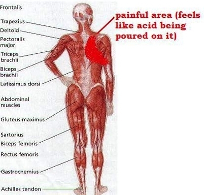
shoulder blade pain | Anatomy System - Human Body Anatomy diagram and chart images
Label Diagram Human Body Stock Illustrations - Dreamstime Download 190 Label Diagram Human Body Stock Illustrations, Vectors & Clipart for FREE or amazingly low rates! New users enjoy 60% OFF. 188,048,419 stock photos online. ... Animal cell structure anatomy infographic diagram. With parts flat vector illustration design for biology science education school book concept microbiology.

Unlabelled Respiratory System Clip Art at Clker.com - vector clip art online, royalty free ...
The following diagram shows cells of onion peel label class ... - Vedantu The following diagram shows human cheek cells, label the parts as observed by you. Answer. Verified. 106.2k+ views. Hint: The diagrams mentioned above are the internal structure of an onion peel and human cheek cells. In order to label them, we need to understand its anatomy and know about various structures present in it. Onion peel is an ...
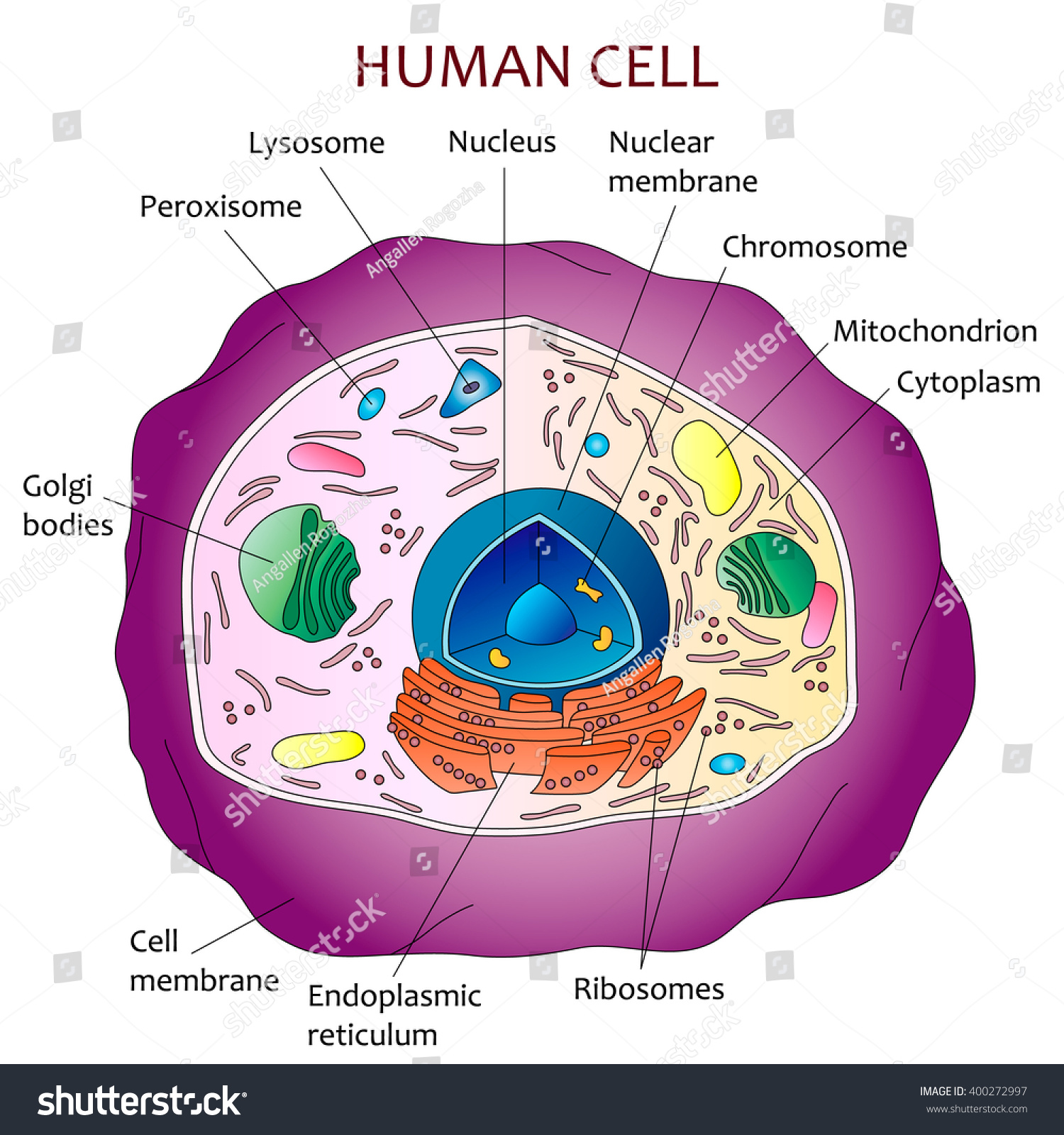
Human Cell Diagram Stock Vector 400272997 : Shutterstock
Labeled Plant Cell With Diagrams - Science Trends The parts of a plant cell include the cell wall, the cell membrane, the cytoskeleton or cytoplasm, the nucleus, the Golgi body, the mitochondria, the peroxisome's, the vacuoles, ribosomes, and the endoplasmic reticulum. Parts Of A Plant Cell The Cell Wall Let's start from the outside and work our way inwards.
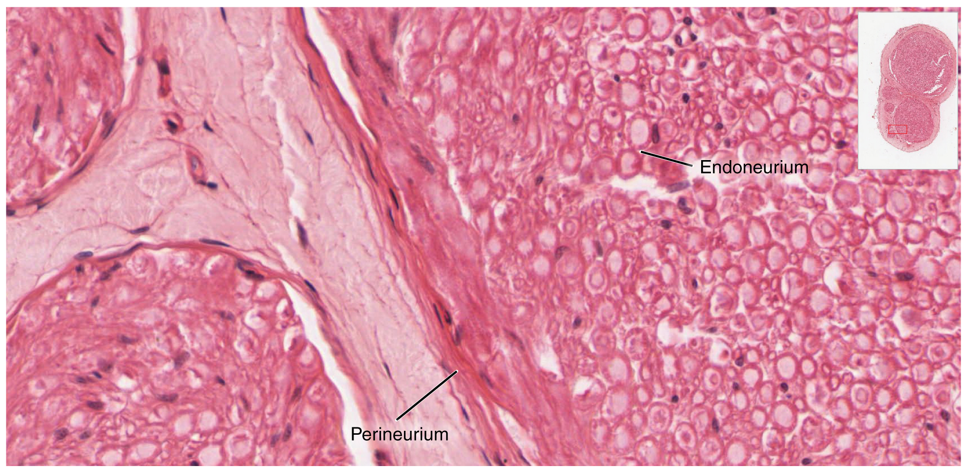
This micrograph shows a magnified view of the nerve. The perineurium and the endoneurium are ...
parts of a human cell | Diagram of the human cell illustrating the ... This diagram of a human skeleton is labeled with 12 major bones, from skull to fibula. D Nicole Science Low Histamine Foods Fodmap Foods Fodmap Recipes Low Fodmap Thyroid Health Gut Health Health Tips Health Fitness Diverticulitis Diet One of the most common digestive challenges I treat is small intestinal bacterial overgrowth (SIBO).

muscular pictures 744×1179 | Anatomy System - Human Body Anatomy diagram and chart images
A multi-omic single-cell landscape of human gynecologic ... Dec 02, 2021 · Deconvolution of regulatory mechanisms that drive transcriptional programs in cancer cells is key to understanding tumor biology. Herein, we present matched transcriptome (scRNA-seq) and chromatin accessibility (scATAC-seq) profiles at single-cell resolution from human ovarian and endometrial tumors processed immediately following surgical resection.

Cell Biology – Cells, Tissues, Organs & Systems | Teaching techniques, Cell biology and Body systems
Human Cell Diagram, Parts, Pictures, Structure and Functions Diagram of the human cell illustrating the different parts of the cell. Cell Membrane. The cell membrane is the outer coating of the cell and contains the cytoplasm, substances within it and the organelle. It is a double-layered membrane composed of proteins and lipids. The lipid molecules on the outer and inner part (lipid bilayer) allow it to ...
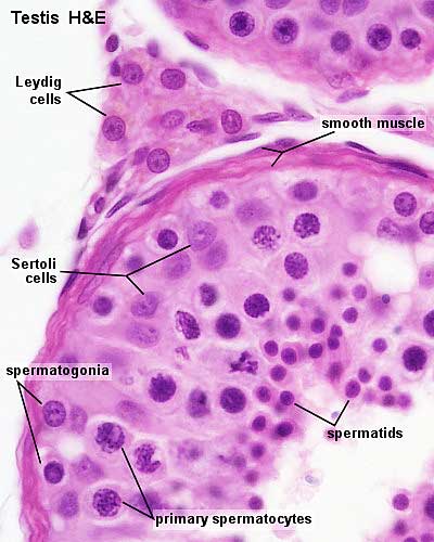
ANAT2341 Lab 1 - Spermatogenesis - Embryology
Blood Cell Diagram Pictures, Images and Stock Photos Realistic vector 3d picture of red blood cells or hemoglobin amount in human blood blood cell diagram stock illustrations. ... Labeled educational blood cancer infographic. Leukemia vector illustration. Labeled educational blood cancer infographic. Bone marrow and abnormal cells disease. Medical diagnosis symptoms and ill treatment list.

Education Chart of Biology for Human Cell Diagram Illustration , #Affiliate, #Biology, #Chart, # ...
03 Label the Cell Diagram | Quizlet Start studying 03 Label the Cell. Learn vocabulary, terms, and more with flashcards, games, and other study tools.

An Annotated Diagram Of A Human Cell
Skeletal System - Labeled Diagrams of the Human Skeleton The skeletal system's cell matrix acts as our calcium bank by storing and releasing calcium ions into the blood as needed. Proper levels of calcium ions in the blood are essential to the proper function of the nervous and muscular systems. Bone cells also release osteocalcin, a hormone that helps regulate blood sugar and fat deposition.

Human Cell Labeled
Human Cell Organelles Labeling Diagram - Quizlet Human Cell Organelles Labeling STUDY Learn Flashcards Write Spell Test PLAY Match Gravity Created by Mackenna_Rios5 Terms in this set (8) Vesicles Transports molecules between organelles and the cell membrane Ribosome Makes Protein Mitochondria Makes ATP Smooth ER Makes lipids and vesicles Lysosomes

Cell Biology – Cells, Tissues, Organs & Systems | Human cell structure, Cell diagram, Eukaryotic ...
A Labeled Diagram of the Animal Cell and its Organelles A Labeled Diagram of the Animal Cell and its Organelles There are two types of cells - Prokaryotic and Eucaryotic. Eukaryotic cells are larger, more complex, and have evolved more recently than prokaryotes. Where, prokaryotes are just bacteria and archaea, eukaryotes are literally everything else.

Interactive cell diagram by Diann Caviness
A pan-tissue DNA methylation atlas enables in silico ... Mar 11, 2022 · For most human tissues and organs, generating DNAm reference profiles for all underlying cell types is very challenging owing to incomplete knowledge of tissue composition and cell-type-specific ...

Plant and Animal Cells......Are they the same?: Process
Human Cells Printables and Diagrams - The Successful Homeschool These cells include: leukocytes, haematids, thrombocytes, ovum, sperm, sarcomeres, enterocytes, neurons, osteocytes, hepatocytes. They will learn the parts of a cell thanks to a labeled diagram. They will get to see what blood looks like under a microscope without needing to own a microscope. They get to color a cell and then label the parts.

Cell-living organisms have cells. This is a diagram of a cell. | physiology:the cel | Pinterest ...
human cell diagram to label Histology basal cell deferens vas label anatomy ansci wisc edu male. Skin histology tissue connective types basic adipose components structure tissues layers system integumentary stain showing dermis dense epidermis irregular embryology. Cell human structure cells cellule nucleus parts anatomy nutshell dna noyau stuff human cell diagram to label
33 Human Cell Diagram To Label - Labels Information List
Cells Diagram | Science Illustration Solutions - Edrawsoft Cells Diagram Symbols Edraw software offers you lots of symbols used in cells diagram like cell structure, paramecium, squamous cell, cell division, bacteria, cell membrane, eggs, sperm, zygote, an animal cell, SARS, tobacco mosaic, adenovirus, coliphage, herpesvirus, AIDS, pollen, plant cell model, onion tissue, etc. Cells Diagram Examples
Post a Comment for "43 diagram of a human cell with labels"