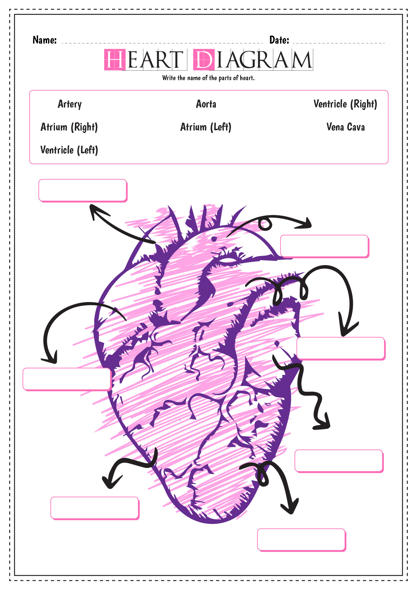
43 human heart diagram without labels
Heart - Wikipedia In humans, other mammals, and birds, the heart is divided into four chambers: upper left and right atria and lower left and right ventricles. [4] [5] Commonly the right atrium and ventricle are referred together as the right heart and their left counterparts as the left heart. [6] anatomyeducation123.z21.web.core.windows.netdiagram of heart without labels Heart diagram without labels clipart parts label cartoon labelling library transparent worksheet clipartkey fireman comprehension reading thump. 12 best images of human brain diagram worksheet. Worksheets heart label human anatomy science sparklebox body diagram ks2 physiology labeling system nursing circulatory diagrams study preschool
commons.wikimedia.org › wiki › File:Diagram_of_theFile:Diagram of the human heart (no labels).svg - Wikimedia Jul 16, 2021 · File:Diagram of the human heart (no labels).svg. From Wikimedia Commons, the free media repository. File. File history. File usage on Commons. Metadata. Size of this PNG preview of this SVG file: 498 × 599 pixels. Other resolutions: 199 × 240 pixels | 399 × 480 pixels | 639 × 768 pixels | 851 × 1,024 pixels | 1,703 × 2,048 pixels | 533 × ...
Human heart diagram without labels
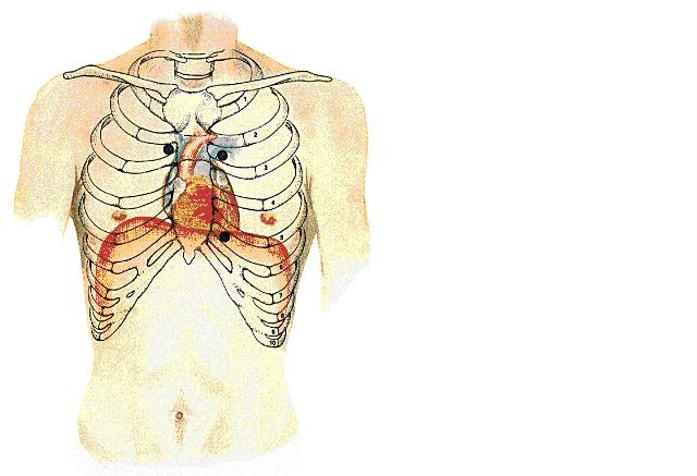
Pericardium—Anatomy and Function - ThoughtCo Pericardium is the membranous sac that surrounds the heart. The pericardium is the fluid-filled sac that surrounds the heart and the proximal ends of the aorta, venae cavae, and the pulmonary artery. The heart and pericardium are situated behind the sternum (breastbone) in a position in the middle of the chest cavity known as the mediastinum. Heart: illustrated anatomy - e-Anatomy - IMAIOS ISSN 2534-5079 Anatomical parts 1 - RCA proximal 1. Basal anterior 10 - Second diagonal 10. Mid inferior 10a - Second diagonal a 11 - Proximal circumflex 11. Mid inferolateral 12 - Intermediate/anterolateral 12. Mid anterolateral 12a - Obtuse marginal a 12b - Obtuse marginal b 13 - Distal circumflex 13. Apical anterior 14 - Left posterolateral 14. Anatomical Line Drawings - Medscape Surface Anatomy - lateral views - male. go to drawing without labels. Surface Anatomy - lateral views - female. go to drawing without labels. Surface Anatomy - Child - anterior view & posterior ...
Human heart diagram without labels. How to Make a DIY Pumping Heart Model - Mombrite Secure with a rubber band or tape. 8. Push both straws through the holes of the balloon. 9. Set the heart model in a tray to catch the "blood.". Make sure to bend the straws downward to avoid projectile blood! 10. Gently press the center of the stretched balloon to pump the blood out of the jar. Circulatory System Diagram - New Health Advisor Coronary circuit mainly consists of cardiac veins including anterior cardiac vein, small vein, middle vein and great (large) cardiac vein. There are different types of circulatory system diagrams; some have labels while others don't. The color blue stands for deoxygenated blood while red stands for blood which is oxygenated. Brigitte Zimmer The human heart is one of the most essential parts of the human body, and getting to know it is imp… Read more How To Draw Human Heart And Label Outline Human Heart Diagram Class 10 Human heart: Anatomy, function & facts | Live Science The human heart is located in the center of the chest - slightly to the left of the sternum (breastbone). It sits between your lungs and is encased in a double-walled sac called the pericardium,...
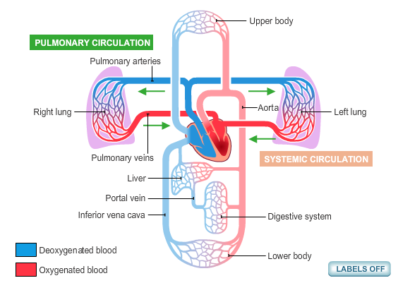
CBSE Class 10 Science Important Biology Diagrams For Last Minute ... The human heart is mainly divided into four parts: two upper parts are called atria, and the lower ones are called ventricles. The ventricles are the chambers that pump blood and atrium are the... Chicken Anatomy 101: Everything You Need To Know Chicken Anatomy of Bone, Legs, and Wings. Bird bones are composed mainly of calcium and phosphorus and a fine web of collagen fibers that are bound tightly together. The skeleton provides support and protection, much as the human skeleton does. 99% of calcium and 80% of phosphorus are stored in the bones. Fetal Circulation Diagram | Fetal Blood Flow & Circulatory System ... The fetal heart like the newborn and adult hearts is made up for four chambers, the right and left atria and ventricles. In newborns and adults, blood flows from the right atrium flows into the ... Circulatory system - Wikipedia Diagram of the human heart showing blood oxygenation to the pulmonary and systemic circulation The heart pumps blood to all parts of the body providing nutrients and oxygen to every cell, and removing waste products. The left heart pumps oxygenated blood returned from the lungs to the rest of the body in the systemic circulation.
Heart - Life processes - Class Notes Heart (1) It is a muscular organ, big as our fist, reddish brown in colour, situated between the 2 lungs in middle of thoracic cavity, surrounded by 2 layered sac. (2) It has different chambers to prevent the oxygen rich blood from mixing with blood containing carbon dioxide. (3) Heart is divided by septa into 2 halves i.e. the right and left. FREE Human Body Systems Labeling with Answer Sheets The free skeletal system labeling sheet includes a fill-in-the-blanks labeling of the main bones on the body. The free respiratory system labeling sheet includes a blank diagram to fill in the trachea, bronchi, lungs, and larynx. The free nervous system labeling sheet includes blanks to label parts of the brain, spinal cord, ganglion, and nerves. Anatomical Planes of Body - The Human Memory The X-axis is going from left to. right, Z-axis from front to back, and Y-axis from up to down. In anatomical. terminology, three references plane are considered standard planes; these. planes differentiate the body anterior and posterior, ventral and dorsal, dexter, and sinister portions. Let me tell you about these standard planes in detail. How the Heart Works: Diagram, Anatomy, Blood Flow The heart is located under the rib cage -- 2/3 of it is to the left of your breastbone (sternum) -- and between your lungs and above the diaphragm. The heart is about the size of a closed fist, weighs about 10.5 ounces, and is somewhat cone-shaped. It is covered by a sack termed the pericardium or pericardial sack.
Label The Heart Diagram - Human Anatomy
How the Heart Works - The Heart | NHLBI, NIH The heart is an organ about the size of your fist that pumps blood through your body. It is made up of multiple layers of tissue. Your heart is at the center of your circulatory system. This system is a network of blood vessels, such as arteries, veins, and capillaries, that carries blood to and from all areas of your body.
Animal Heart Diagram Labeled - ClipArt Best
Female Anatomy: Labeled Diagrams of the Reproductive System Female anatomy refers to the internal and external structures of the reproductive and urinary systems. Reproductive anatomy aids with sexual pleasure, getting pregnant, and breastfeeding a baby. The urinary system helps rid the body of toxins through urination (peeing). The Female Reproductive System. Some people are born with internal or ...

Heart Diagram | Anatomy Of Heart | Different Parts Of The Heart
Parts of the Heart & Blood Flow | Diagram & Overview - Study.com Diagram of Heart Valves The first valve is the tricuspid valve. It gets its name because it has three cusps that anchor it down into the right ventricle. The tricuspid valve is located at the exit...

heart diagram no labels
Heart Labeling Quiz: How Much You Know About Heart Labeling? Create your own Quiz Here is a Heart labeling quiz for you. The human heart is a vital organ for every human. The more healthy your heart is, the longer the chances you have of surviving, so you better take care of it. Take the following quiz to know how much you know about your heart. Questions and Answers 1. What is #1? 2. What is #2? 3.

Human heart diagram, Heart diagram, Heart printable
Left Coronary Artery: Anatomy, Function, and Significance The left coronary artery and its branches play a crucial role in ensuring that the muscles of the heart, itself, are supplied with oxygenated blood. 1 Specifically, it provides the majority of supply to the ventricles (the lower chambers of the heart) as well as the left atrium and atrial appendage, the pulmonary artery, and aortic root.

Label the heart — Science Learning Hub
Path of Blood Through the Heart - New Health Advisor Basics Parts of the Heart. Understanding the function of the heart is helpful to learn more about its anatomy. Here are the basic parts of the heart: 1. Right Atrium. The heart can be divided into right and left halves, as well as into the upper and lower chambers. There are two upper chambers called atria and two lower chambers called ventricles.
13+ Heart Diagram Templates – Sample, Example, Format Download | Free & Premium Templates
anatomyeducation.z21.web.core.windows.net › humanhuman heart diagram without labels Free Unlabeled Heart Diagram, Download Free Clip Art, Free Clip Art On. clipart-library.com. heart diagram human simple drawing blank circulatory system labels sketch unlabeled easy blood label worksheet anatomy flow circulation labelling library.
Circulatory System Diagram | New Health Advisor
Blank ear diagrams and quizzes: The fastest way to learn - Kenhub It helps you to memorize the names and their locations, which in turn will aid you to remember their functions. Below, you can download both the blank ear diagram to make some notes, and then try labeling the ear using the unlabeled ear diagram. Good luck! DOWNLOAD PDF WORKSHEET (BLANK) DOWNLOAD PDF WORKSHEET (LABELED)

File:Diagram of the human heart uk.svg - Wikimedia Commons
Cardiovascular system: Diagrams, quizzes, free worksheets - Kenhub In this diagram of the cardiovascular system, you can see labeled structures. Spend a few minutes analysing the diagram, and trying to connect the location of the structures with what you've learned in the video. Once you think you've got a solid idea, it's time to try our cardiovascular system labeling quiz.
11 Best Images of Blank Heart Diagram Worksheet With Word Bank - Label Heart Diagram Worksheet ...
Anatomy of The Human Ribs - With Full Gallery Pictures! The Anatomy of the Human Ribs (costae) are one of the integral parts of the chest wall; they make up the lateral part of our body, its anterior and posterior wall and they entirely build the lateral parts of the chest wall. The anatomy of the human ribs is made up of 24 ribs. These ribs are parted in 12 pairs (each on the left and right side of ...

mypicsainmarin: heart diagram with labels
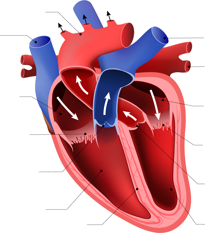
in.pinterest.com › pin › human-heart-diagram-withoutHuman Heart Diagram Without Labels | Human heart diagram ... Credits: All photography, text, and labels by Rob Swatski, Assistant Professor of Biology, Harrisburg Area Community College - York Campus, York, PA. Email: rjswatsk@hacc.edu This work bears an Attribution-Noncommercial Share Alike Creative Commons 3.0 license. All other diagrams and illustrations used in…
a Lebelled Diagram of the Heart : Anatomy and physiology Heart - Cancer / Asbestos Cancer ...
Human Heart Drawing - How To Draw A Human Heart Step By Step Step 1ShareShare on Pinterest Share on Facebook. For this first step of our guide on how to draw a human heart, we will start with some outlines for the heart. We will start with the aorta, which is the pipe-like part at the top of the heart. Use some curved lines for this aorta with a small oval shape at the tip of it.

Human Heart Diagram Without Labels | Human heart diagram, Heart diagram, Human heart
› pin › 147633694010620242Human Heart Diagram Without Labels | Human heart diagram ... Jessica Muratorri. Vet tech. Cranial Anatomy. This exhibit depicts the anatomy of the inferior skull including: the foramen magnum, occipital condyles, mastoid process, styloid process, mandibular fossa, palatine bone, sphenoid bone, carotid canal, and the jugular fossa. E.
Post a Comment for "43 human heart diagram without labels"